Sample No. 0216
Sublingual Lesions in Cats: Ranulas, Eosinophilic Disease and Other Causes
A swelling beneath a cat’s tongue can have several causes. Veterinary examination and tissue testing can help distinguish them and guide care.
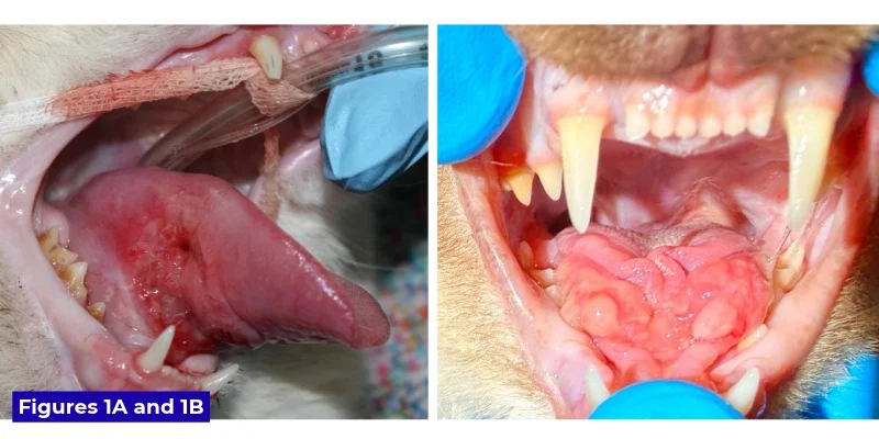
A mass or swelling beneath a cat’s tongue deserves prompt veterinary attention, but its appearance alone may not reveal the cause. Squamous cell carcinoma is a serious possibility, particularly in an older cat with bloody drool, reduced appetite or trouble swallowing. Yet ranulas, eosinophilic granulomas and less common problems can look similar. The practical lesson is to keep several possibilities in mind and use examination and, when needed, tissue analysis to establish a diagnosis.
When saliva collects beneath the tongue
A ranula is a swelling beneath the tongue caused by saliva escaping from a damaged salivary gland or duct. The name comes from the Latin word rana, meaning frog; the smooth swelling can resemble a frog’s underside. It may appear on one side or both, and is usually not inflamed at first.
The openings of the mandibular and sublingual salivary ducts, called caruncles, sit near the midline under the tongue. Damage to these openings or to the ducts leading from the glands can allow saliva to collect in nearby tissue. If the swelling becomes large, contact with the teeth may cause additional injury. Some ranulas settle without intervention, while others require veterinary surgery to remove the mandibular and sublingual glands.
Recent oral surgery can complicate the picture. When a procedure to remove part of the jaw requires cutting and tying off salivary ducts, temporary swelling beneath the tongue may resemble a ranula. These postoperative “pseudoranulas” are described as resolving within a few days. A small 1978 experiment found that surgical damage to a duct or gland did not consistently produce a salivary cyst in dogs. The finding does not rule out postoperative ranulas: the author also describes seeing one after ducts were cut during cancer surgery.
Eosinophilic disease can imitate a mass
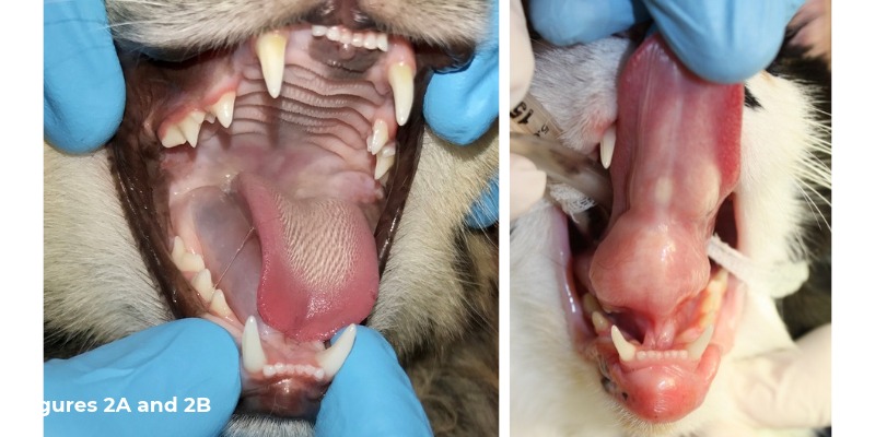
Eosinophilic granuloma is often associated with ulcers on a cat’s upper lip, sometimes called rodent ulcers. The condition can also cause swelling without that familiar appearance, including beneath the tongue. A sublingual lesion may look smooth and fluid filled, like a ranula, or firm, ulcerated and irregular, raising concern for cancer.
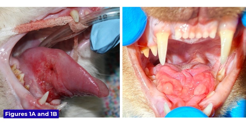
Other signs can help shape the veterinary assessment. A cat with a sublingual eosinophilic lesion may also have a swollen lower lip. In one reported case, a one-year-old Ragdoll named Milo had a very firm, wartlike, ulcerated swelling beneath the tongue and a history of lower-lip swelling. Tissue analysis identified eosinophilic granulomatous glossitis with ulceration. The appearance had raised concern for squamous cell carcinoma.
Because the lesion was severe and at risk of being traumatized by the teeth, the veterinary team began steroid treatment while awaiting the biopsy result. The swelling had decreased substantially within a week and was nearly gone after two weeks. A dermatology consultation was recommended to investigate possible allergic causes. The case illustrates why appearance and history can guide the investigation, but tissue testing may be needed to confirm what a lesion is.
Look for irritation or a hidden foreign body
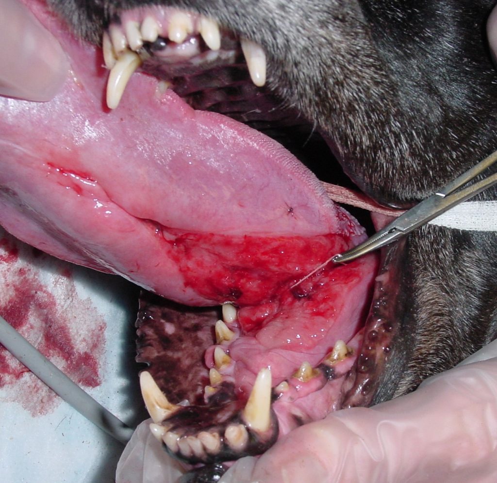
A linear foreign body caught around the lingual frenulum—the tissue under the tongue that helps anchor it—can cause marked swelling and redness beneath the tongue, as well as swelling of the tongue itself. This problem is seen most often in young cats, though dogs can be affected. The source describes a ten-year-old dog whose chewing on a golf ball’s inner twine left it wrapped around the base of the tongue, an unusual age for this presentation.
Chronic rubbing can also produce a broad patch of granulation tissue beneath the tongue. One possible source of ongoing irritation is a rough tooth surface associated with tooth resorption. A veterinarian can examine the mouth for a foreign object, dental injury or other source of repeated contact.
Get a new or changing lesion checked
Bloody drool, difficulty swallowing, reduced appetite, ulceration or a rapidly enlarging swelling are reasons to arrange veterinary care promptly. A visible lesion may be hard to assess at home, and trying to inspect or manipulate a painful area can cause further distress. Your veterinarian can examine the mouth and decide whether imaging, sampling or biopsy is appropriate. If a swelling appears after oral surgery, contact the treating team so they can assess whether it fits the expected recovery or needs attention.
Note any changes in eating, drooling, lip swelling or the lesion’s size and appearance, and share when they began. These observations can help the veterinary team consider the history alongside the examination. Because several conditions can resemble one another beneath the tongue, a careful assessment—and tissue analysis when indicated—can help direct the next steps.